The initial test for couples seeking treatment for infertility or who wish to have children is a “sperm analysis.” Sperm analysis is the most commonly used test for evaluating male infertility and is considered a standard test. In addition, there are some basic and established tests that have been used for many years to evaluate male infertility. These include chromosome analysis, Y microdeletion testing, and genetic analyses of genes such as the CFTR gene, KAL1 gene, androgen receptor (AR) gene, LH gene, GnRH gene, and SRD5A2 gene, which are selected based on the patient's findings.
Today, new generation tests have been introduced in addition to these tests. These are particularly valuable tests for men with azoospermia, a condition in which there are no sperm cells. This is because there are many known and unknown causes of azoospermia. To overcome azoospermia, it is first necessary to accurately identify the cause, as only then can the appropriate treatment be planned and the problem resolved. Otherwise, the number of unhappy and exhausted patients who undergo the same procedure repeatedly will continue to increase.
These new generation tests:
WES-based Male Infertility Panel
Here, more than 300 gene scans are performed to identify the cause. Thanks to these tests, we can clearly identify the cause, make changes to our treatment protocol based on the cause, or, unfortunately, sometimes we have to tell our patient that there is no hope.
Spermatogenesis Gene Expression Analysis
Haploid (meiosis) panel from semen sample; the first and most critical stage of sperm production is meiosis, which reduces the number of chromosomes by half. This test determines whether the genes that control meiosis are active (expression levels). For this purpose, we can determine whether meiosis is present by testing PCNA, Lim15, ESX1, TEX101, and other genes. Other genes studied include UBQLN3, FAM71F1, CAPN11, SPATA3, GGN, and SPACA4. The most important advantage of this test is that it allows us to obtain information about sperm production in the testicles from a semen sample without performing a surgical procedure such as TESE. Some other important advantages of this test are listed below:
-
Spermatogenesis testicles (meiosis) whether it is active or not
detection from semen sample (non-invasive)
-
Unnecessary/Unsuccessful Reducing the TESE rate
-
Determining the optimal timing for TESE
-
Enabling treatment follow-up and determining whether the patient responds to treatment
is that it can be tracked accurately.
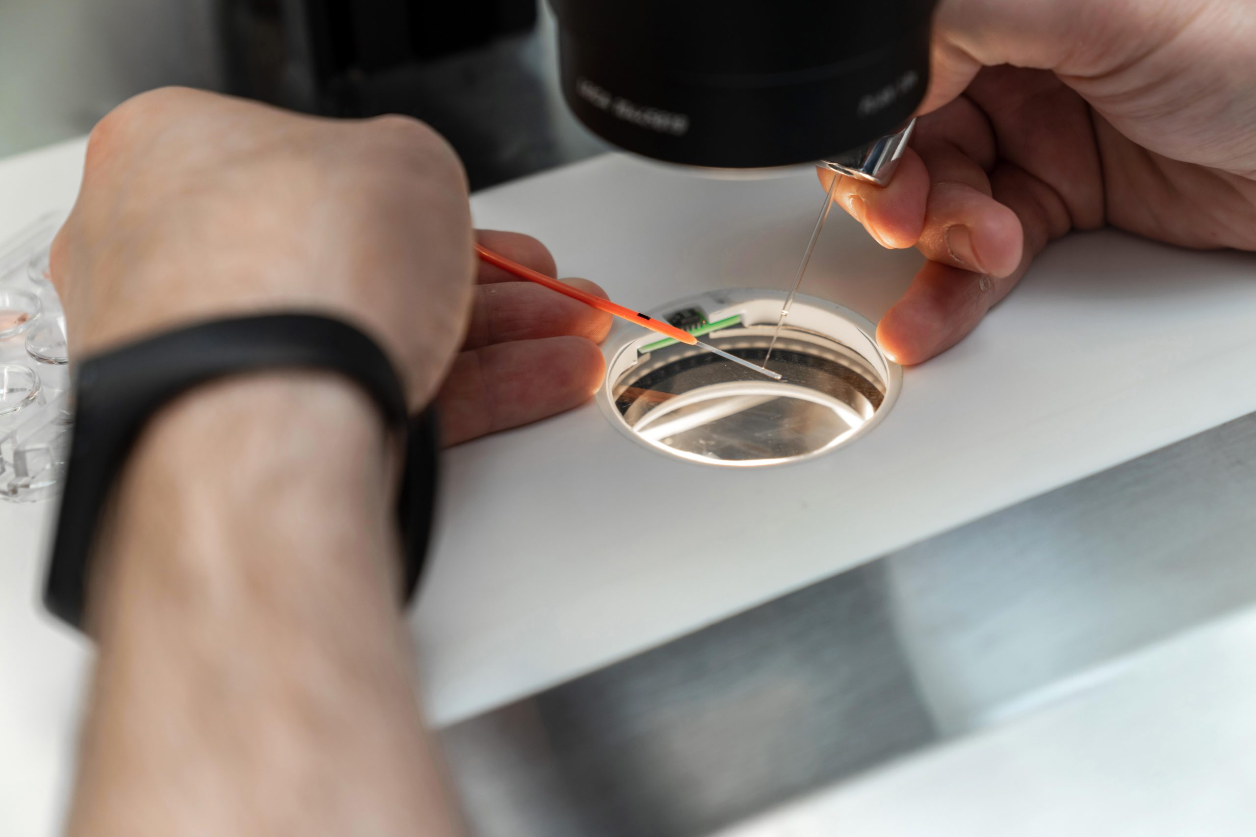
Flow Cytometry Analysis
Flow cytometry analyses are extremely reliable and high-tech tests approved by the FDA. With flow cytometry testing, we can detect the presence of certain cells or organic structures in the environment and even determine the percentage of these cells. This method uses antibodies to detect the presence and quantity of cells we wish to identify by reading them with a laser, enabling us to obtain highly specific and reliable results. Moreover, this method allows for the reading and control of hundreds of thousands of cells. For example, this method can detect sperm cells in a semen sample from a patient who was previously diagnosed with azoospermia, meaning no sperm were visible under a standard microscope. Other important benefits of this method are listed below:
-
Determination of the presence of sperm cells in semen
-
Determination of sperm DNA damage percentage
-
Determination of oxidative stress (ROS) levels in semen
-
Determination of apoptosis and preapoptosis levels in Semende


These new generation tests are not only used in cases of azoospermia. Sometimes, even if the spermogram test is normal, we still need new generation tests. For example, if an embryo does not develop beyond the third day or does not develop into a blastocyst, the problem may be sperm-related; even if the male partner's sperm count, morphology, and motility are normal, the problem may still be caused by the male partner. New-generation flow cytometry tests that show sperm DNA or membrane potential can reveal the source of the problem and enable pregnancy to be achieved following treatment of the male partner.
Artificial Oocyte Activation (AOA) and Piezoelectricity
One of the most critical stages of fertilization after the sperm cell enters the egg cell is oocyte activation. Normally, this stage begins physiologically following the fusion of the sperm and oocyte membranes, triggered by factors released from the sperm head (Sperm Oocyte-Activator Factor – SOAF) through the release of calcium (Ca+2 oscillation) via a molecule called inositol trisphosphate (IP3). In IVF applications, we sometimes encounter situations where eggs collected from the female partner do not fertilize after sperm injection (ICSI). This is called fertilization loss. If the egg does not activate physiologically after ICSI, it may be possible to activate the egg using artificial methods. Among these methods are “PLCzeta” microinjection and “ionophore treatment,” but these have been observed to increase sperm DNA damage rates and cause side effects such as “premature acrosome reaction” (beginning before sperm integration into the oocyte), or in many cases, sufficient success has not been achieved. Recently, the “Piezoelectric Activation” method has been introduced among artificial activation methods for egg fertilization. The mechanism of piezoelectric application involves stimulating calcium channel proteins within the egg's cell membrane through electromagnetic current, followed by triggering calcium influx (calcium oscillation) upon the opening of these channels. This method is becoming increasingly widespread.